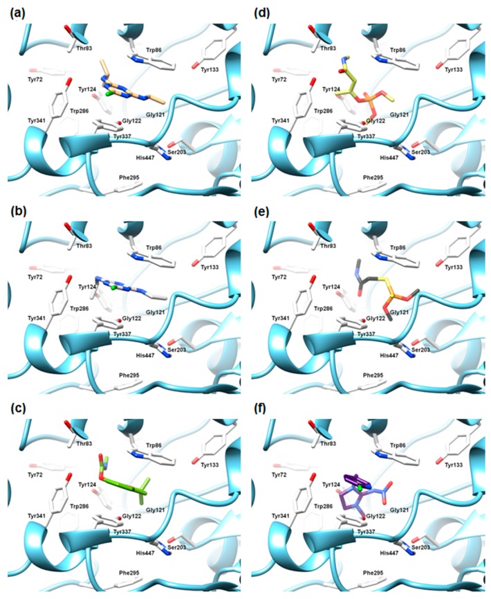
Figure 4.
The SB alignment of atrazine (a); simazine (b); carbofuran (c); monocrotophos (d); dimethoate (e); imidacloprid (f) into the mAChE active site. The enzyme ribbons are presented in blue, the active site amino acids are depicted in white. For the purpose to clarify, the hydrogen atoms are omitted. The graphic was generated using the UCSF Chimera software (Resource for Biocomputing, Visualization, and Informatics (RBVI), University of California, San Francisco, CA, USA).

