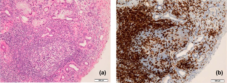
Fig. (2).
Ectopic GC formation in the salivary gland tissue from SS patients. (a) Inflammatory lesions including CG in the lip biopsy tissue from a SS patient is shown by histological staining with hematoxylin and eosin. A lot of lymphocytes infiltrate extensively in the salivary gland tissue with destruction of acinar cells. (b) CD3+ T cells in lip biopsy tissue from a SS patient are shown by immunohistochemistry. Scale bar: 200 μm.

