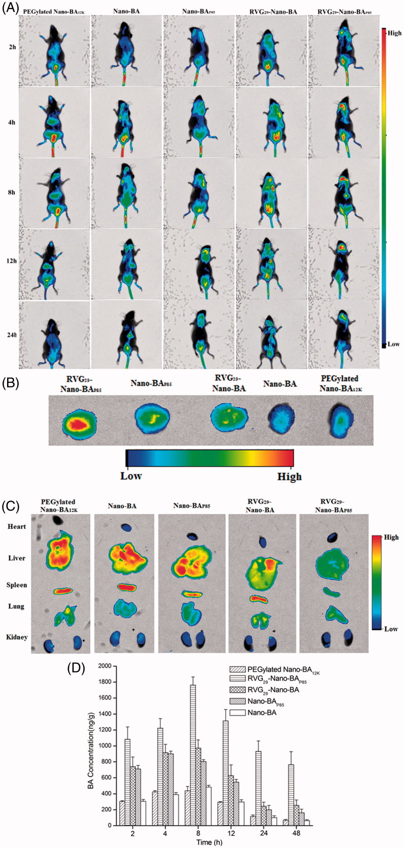
Figure 5.
The in vivo noninvasive images of time-dependent whole body imaging of PM mice co-infected with S. pneumonia ATCC 49619 and S. pneumonia 16167 after i.v. injection of DiR-loaded RVG29-Nano-BAP85, RVG29-Nano-BA, Nano-BAP85, Nano-BA, and PEGylated Nano-BA12K (A). The ex vivo optical images of the brain (B) and other organs (C) of the PM mice co-infected with S. pneumonia ATCC 49619 and S. pneumonia 16167 after i.v. injection of DiR-loaded RVG29-Nano-BAP85, RVG29-Nano-BA, Nano-BAP85, Nano-BA, and PEGylated Nano-BA12K. Quantitative analysis for accumulation in the brain of BA in the PM mice co-infected with S. pneumonia ATCC 49619 and S. pneumonia 16167 at different times after intravenous administration of RVG29-Nano-BAP85, RVG29-Nano-BA, Nano-BAP85, Nano-BA, and PEGylated Nano-BA12K at a dose of 30 mg/kg, respectively (D).

