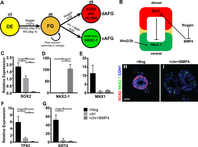
Figure 2:
Anterior foregut spheroids have esophageal-respiratory competence.
(A) Schematic depicting experimental protocol to pattern AFG spheroids along the dorsal-ventral axis. (B) Current simplified model of the cues guiding dorsal-ventral patterning of the AFG of mouse and frog embryos. (C-G) qPCR analysis of 3-day-old spheroids (day 9) treated for 3 days with Noggin, untreated (ctrl), or chiron and BMP4 (10ng/mL) using dorsal markers SOX2 and MNX1 (C+E), the respiratory marker NKX2–1 (D), ΔN splice variant of TP63 (F), and the stratified squamous epithelium marker KRT4 (G). (H-I) IF staining for SOX2, NKX2–1, CDH1, and nuclei (DAPI) in Noggin (H) versus chiron+BMP4 (I) treated spheroids. Scale bar = 25μm. See quantification and statistical analysis section for details. See also Figure S3.

