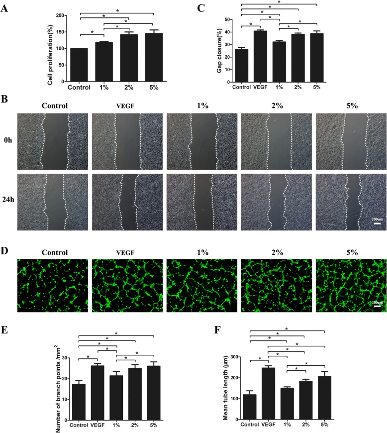
Fig. 6.
FE promotes endothelial cell proliferation, migration, and tube formation. a HUVECs treated with FE at indicated concentrations. Cell proliferation assessed using cell counting kit, and percentage of optical density values relative to control calculated. b HUVEC migration evaluated using cell migration assay. c Percentage of gap closure (24 h) quantified. d HUVECs added to solidified Matrigel in a serum-free medium in presence or absence of FE. VEGF 20 ng/ml used as positive control. After 6-h incubation, endothelial cell tube formation stained using Calcein-AM and assessed by fluorescence microscopy. e Assessment of number of branch points/mm2 in each group. f Quantification of mean tube length. *p < 0.05. VEGF vascular endothelial growth factor

