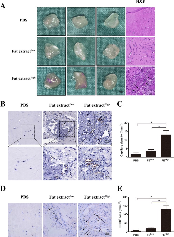
Fig. 7.
Matrigel plug assay shows that FE promotes angiogenesis in vivo. a PBS and FE of low and high concentrations mixed with Matrigel and injected subcutaneously into dorsal region of nude mice. Matrigel plugs harvested at 1 week post implantation. Left panel: gross morphology of Matrigel plugs. Right panel: hematoxylin–eosin (H&E) staining of paraffin sections of explanted plugs. Tissue with more blood vessels observed in FEHigh plugs. Arrows indicate formed blood vessels. b Immunostaining of Matrigel plug with CD31 antibody. FEHigh group showed more CD31+ blood vessels. c Quantification of CD31+ capillary density. Significantly higher blood vessel density measured in FEHigh-treated group. d Immunostaining of mouse macrophage marker CD68 on Matrigel plugs. FEHigh group showed more CD68+ cells. e Quantification of CD68+ cells. Significantly higher number of CD68+ cells observed in FEHigh-treated group. *p < 0.05. PBS phosphate-buffered saline

