Fig. 1.
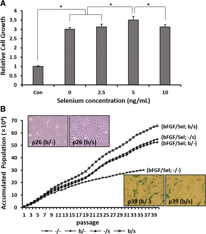
In vitro expansion of AF-MSCs treated with bFGF and selenium. a Relative growth rates of AF-MSCs after 3 days of exposure to various concentrations of selenium (0, 2.5, 5, and 10 ng/mL) under bFGF (4 ng/mL) supplementation. The growth rates were determined by crystal violet assay. Error bars represent the mean ± SD of three independent experiments performed in triplicate. *p < 0.05. b Proliferation of AF-MSCs cultured with bFGF (4 ng/mL) and selenium (5 ng/mL), alone or in combination, until they reached senescence. The inset images show the morphology of AF-MSCs in the proliferation stage (top images) and large and flattened AF-MSCs expressing β-gal as a marker of cellular senescence (bottom images), respectively

