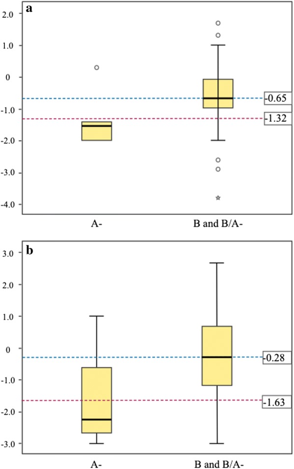
Fig. 3.

Box plots showing the distribution of absolute changes in haemoglobin concentration of G6PD deficient and non-deficient subjects on the 3rd day (a), and the 7th day (b), relative to basal levels before primaquine treatment on day 1. The dotted line indicates median. The Y axis indicates the absolute loss of haemoglobin (g/dL). Genotypes A (deficient) and B (normal) are indicated in the X axis
