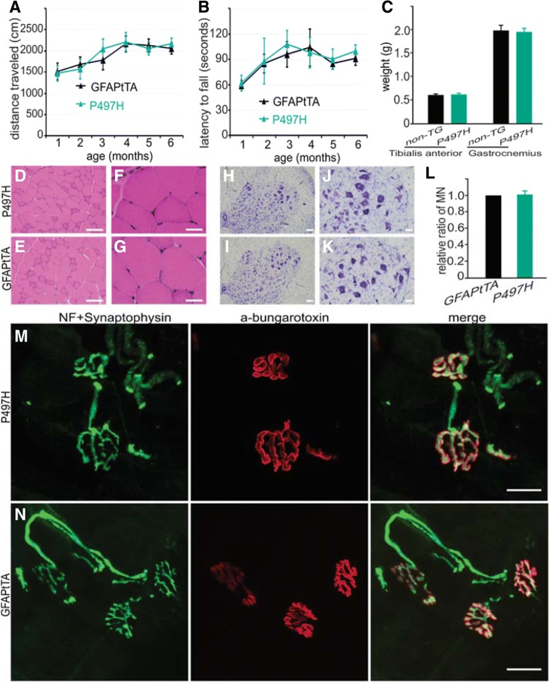
Fig. 8.
No motor phenotypes in GFAPtTA/UBQLN2P497H rats. a-b Results of beavioral tests in GFAPtTA/UBQLN2P497H bigenic (P497H) and GFAPtTA single transgenic rats. c The weights of tibialis anterior and gastrocnemius muscles at 6 months old in P497H and GFAPtTA female rats. The data are reported as the mean ± standard deviation (n = 4). d-g H&E staining shows the structures of gastrocnemius muscle in both P497H and GFAPtTA rats; h-l Cresyl violet staining shows the motor neurons in the ventral horn of the spinal cord. The quantification of motor neurons in the L3–5 spinal cord shows that there is no statistical difference between P497H and GFAPtTA rats (n = 4). m-n Confocal images show the structures of neuromuscular junctions (NMJ) in gastrocnemius muscles. The sections were stained with the presynaptic neuronal marker neurofilament (NF) and synaptophysin together with α-bungarotoxin to show the post-synaptic structures. Scale bars: 100 μm (d, e, h, i), 50 μm (j, k), 30 μm (f, g), 20 μm (m, n)

