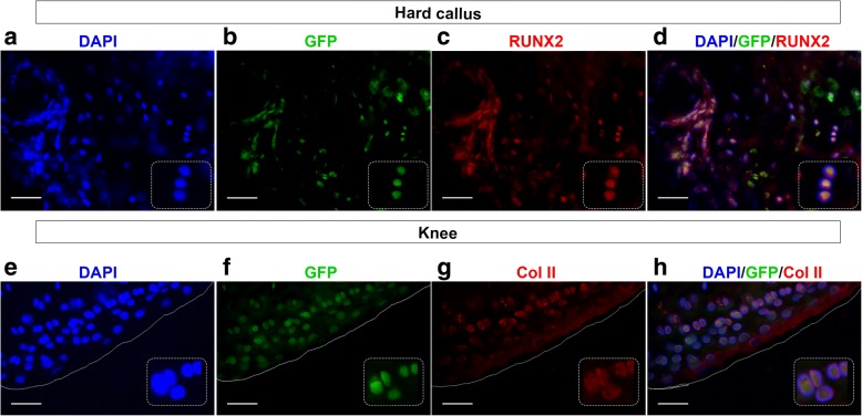
Fig. 3.
GFPpos BST2pos-CH cells differentiate into tissue-specific cells. BST2pos-CH cells, isolated from the peripheral blood of GFP-transgenic naive mice and intravenously injected in syngeneic WT mice are able to engraft and differentiate in bone and cartilage tissue-specific cells, 24 days after. a–d Serial fluorescence images showing signals from DAPI (blue) (a), endogenous GFP (green) (b), and anti-Runx2 (red) (c) in the hard callus of fractured cell-injected mice. Overlap between DAPI, GFP, Runx2 is shown in panel d. White inset boxes in the panels show a higher magnification of representative cells co-expressing DAPI, GFP, and Runx2 signals. e–h Serial fluorescence images showing signals from DAPI (blue) (e), endogenous GFP (green) (f), and anti-Col II (red) (g) in knee region of fractured cell-injected mice. Overlap between DAPI, GFP, and Col II is shown in panel h. White inset boxes in the panels show a higher magnification of representative cells co-expressing DAPI, GFP, and Col II signals. Magnification × 40, scale bar 50 μm

