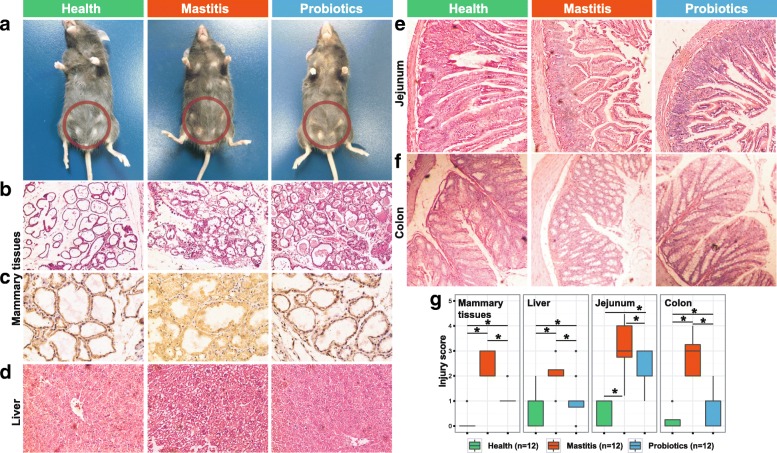
Fig. 2.
Histological analysis of mouse tissues after FMT and probiotics intervention. a Pathological changes in mammary gland surface, where two abdominal mammary glands were swelling in the mastitis group of mice on day 25 after FMT. Breast of mice was highlighted by red circles. b Representative photomicrographs of hematoxylin-eosin stained mammary gland tissue (× 200 magnification). c CD45 immunohistochemical staining sections at × 400 magnification. d–f Representative photomicrographs of hematoxylin-eosin stained liver (× 200), jejunum (× 100), and colon tissue (× 100). g The injury score of mammary gland, liver, jejunum, and colon

