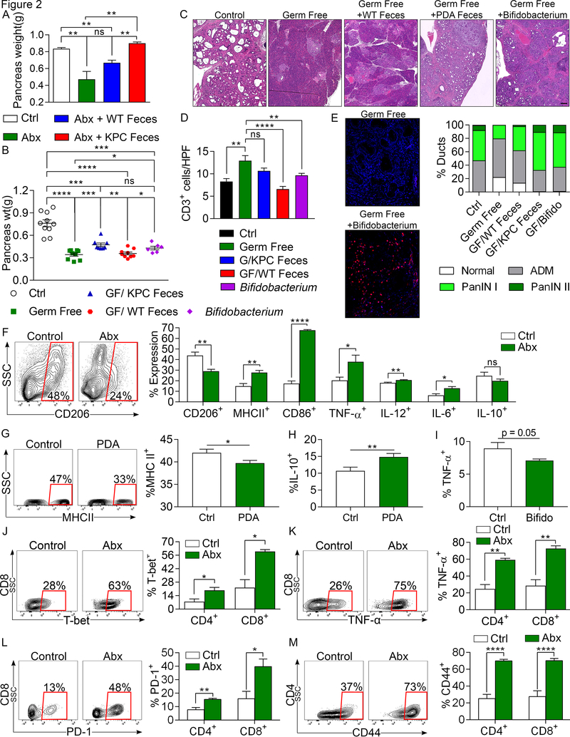
Figure 2. The microbiome in PDA-bearing hosts promotes tumor-progression and intra-tumoral immune suppression.
(A) KC mice treated with an ablative oral antibiotic regimen for 8 weeks were repopulated with i) feces from 3-month old WT mice, ii) feces from 3 month-old KPC mice, or iii) sham-repopulated (vehicle only). Mice were sacrificed 8 weeks later and pancreas weights from each cohort were compared to each other and to age-matched control KC mice that were not treated with antibiotics (n=3–4/group). This experiment was repeated three times. (B-D) The gut microbiome of germ-free 6-week-old KC mice were repopulated with feces from 3-month-old WT or KPC mice, B. pseudolongum, or sham-repopulated. Mice were sacrificed 8 weeks later. (B) Tumor weights were measured. Each point represents data from a single mouse. (C) Representative H&E-stained sections of pancreata are shown compared with age-matched non-germ free controls (scale bar =100μm). Ductal histology was quantified. (D) CD3+ T cell infiltration was determined by IHC. All repopulation experiments were repeated 3 times. (E) The gut microbiome of germ-free 6-week-old KC mice were repopulated with B. pseudolongum or sham-repopulated (n=5/group). Colonization of pancreata with B. pseudolongum was confirmed using FISH at 8 weeks. This experiment was repeated twice. (F) Control and oral antibiotic-treated WT mice were orthotopically implanted with KPC-derived tumor cells. Gr1–CD11b+F4/80+ macrophages were gated and assessed for expression of CD206, MHC II, CD86, TNF-α, IL-12, IL-6, and IL-10 (n=5/group). Macrophage profiling experiments were repeated more than 5 times (G, H) Splenic macrophages from untreated mice were harvested and cultured in vitro with cell-free extract from gut bacteria of control or KC mice. After 24h, macrophages were analyzed for expression of (G) MHC II and (H) IL-10 (n=18/group). (I) Splenic macrophages were cultured in vitro with cell-free extract from B. pseudolongum or with PBS. After 24h, macrophages were analyzed for expression of TNF-α. Macrophage polarization experiments were repeated 3 times in replicates of 5. (J-M) Control and oral antibiotic-treated WT mice were orthotopically implanted with KPC-derived tumor cells. CD4+ and CD8+ T cells were gated and tested for expression of (J) T-bet, (K) TNF-α, (L) PD-1, and (M) CD44. Representative contour plots and quantitative data are shown. Immune-phenotyping experiments were repeated more than 5 times (n=5/group; *p<0.05, **p<0.01, ***p<0.001, ****p<0.0001).

