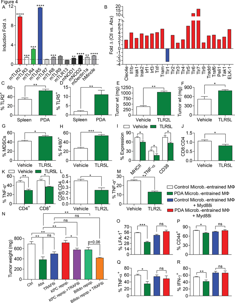
Figure 4. The PDA microbiome induces immune suppression via differential TLR activation.
(A) Cell-free extract from gut bacteria derived from 3 months old WT or KC mice (n=3) were tested for activation of a diverse array of PRR-specific HEK293 reporter cell lines. (B) Orthotopic KPC tumors were harvested on day 21 from control and oral antibiotic-treated WT mice. PRR-related gene expression in PDA was determined using a PCR array and performed in duplicate. Data indicates fold change in gene expression for control compared to antibiotic-treated groups. This array was repeated twice. (C) Expression of TLR2 and (D) TLR5 were tested in spleen and PDA-infiltrating macrophages from orthotopic KPC tumors (n=5). These experiments were repeated twice. (E) WT mice were orthotopically implanted with KPC-derived tumor cells and serially treated with TLR2 (Pam3CSK4) or (F) TLR5 (Flagellin) ligand or vehicle. Tumor growth was determined at 3 weeks (n=3–5/group). These experiments were repeated twice. (G-K) WT mice were orthotopically implanted with KPC-derived tumor cells and serially treated with TLR5 ligand. Tumors were harvested at 3 weeks and (G) the fraction of Gr1+CD11b+ MDSC and (H) F4/80+Gr1–CD11b+ TAM infiltration was determined by flow cytometry. (I) Expression of MHC II, TNF-α, and CD38 on TAMs were determined. (J) The CD8/CD4 T cell ratio was determined as was (K) TNF-α expression on CD4+ and CD8+ T cells (n=3–5/group). (L, M) WT mice were orthotopically implanted with KPC-derived tumor cells and serially treated with TLR2 ligand. Tumors were harvested at 3 weeks and (L) the CD8/CD4 T cell ratio and (M) TAMs expression of TNF-α were determined. These experiments were repeated twice (n=3–5/group). (N) WT mice pre-treated with an ablative oral antibiotic regimen or vehicle for 6 weeks were repopulated with i) feces from 3 month-old KPC mice (n=12), ii) B. pseudolongum (n=6), or iii) sham-repopulated (n=4). Mice were challenged with orthotopic KPC cells. Cohorts were additionally treated serially with a TRAF6 inhibitor or control. Treatments were started at the time of tumor implantation and continued until sacrifice at 21 days. Quantitative analysis of tumor weights are shown. (O-R) Splenic macrophages were entrained with extract from the gut microbiome of either WT or KC mice in the context of MyD88 inhibition or control. Macrophages were then used in αCD3/αCD28-based T cell stimulation assays. CD4+ T cell activation was determined by expression of (O) LFA-1, (P) CD44, (Q) TNF-α, and (R) IFN-γ. This experiment was repeated five times in 3–4 replicates per group with similar results (*p<0.05, **p<0.01, ***p<0.001, ****p<0.0001).

