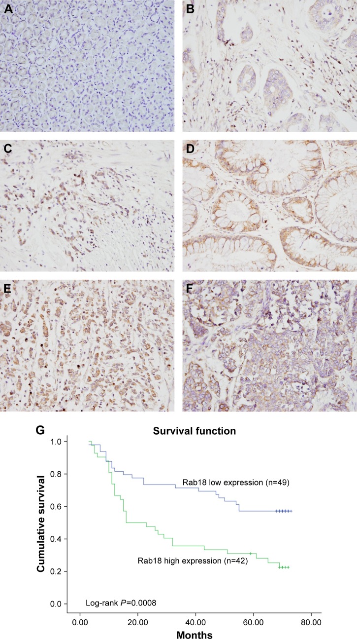
Figure 1.
Expression of Rab18 in gastric cancers.
Notes: (A) Negative Rab18 staining in normal gastric tissues. (B) Weak cytoplasmic Rab18 staining in a sample of papillary gastric carcinoma. (C) Positive Rab18 staining in a sample of mucinous adenocarcinoma. (D) Positive Rab18 expression in a sample of tubular adenocarcinoma. (E) Positive staining in a sample of signet-ring cell carcinoma. (F) Rab18 staining in a sample of neuroendocrine carcinoma. (G) High Rab18 expression correlated with poor patient survival (log-rank test, P=0.0008).

