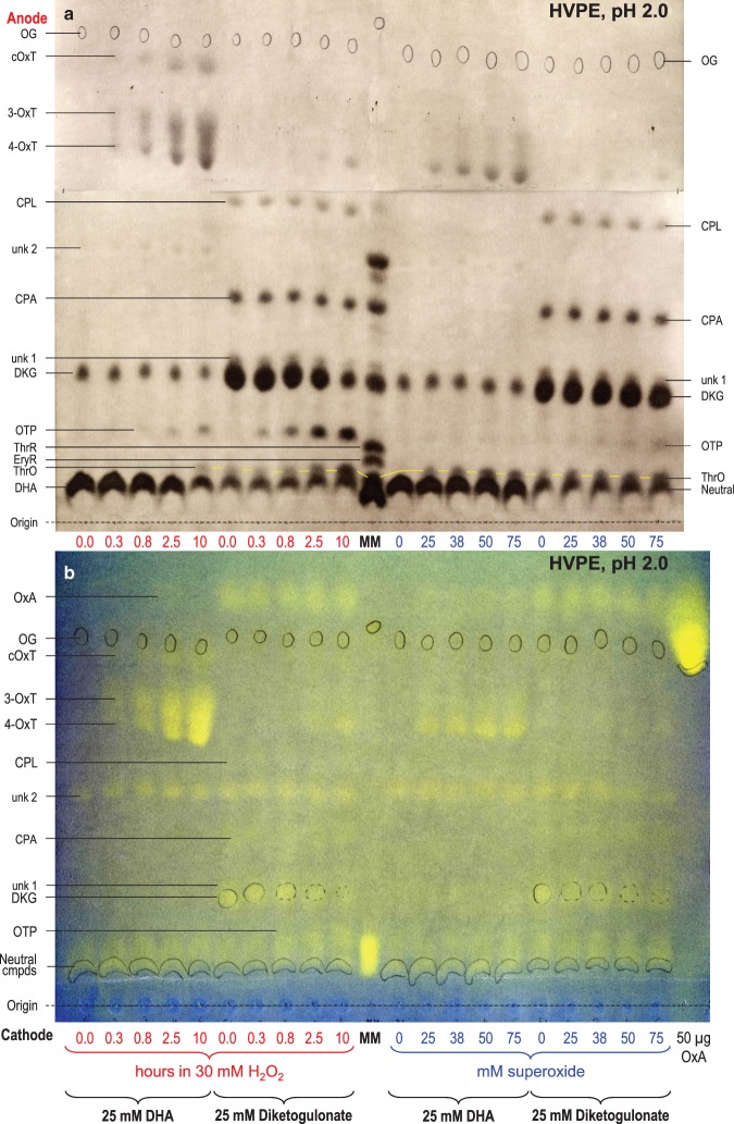
Figure 4. Reaction of dehydroascorbic acid and diketogulonate with H2O2 and superoxide: electrophoresis at pH 2.0.
DHA or DKG (25 mM) was incubated with 30 mM H2O2 for 0–10 h (left half of each electrophoretogram), or with 0–75 mM KO2 in the presence of catalase for a few seconds (right half). All reaction mixtures were buffered with 200 mM acetate (Na+), pH 4.7. Samples were electrophoresed for 60 min at pH 2.0. (a) 10 µl sample run at 2.5 kV with AgNO3 staining. (b) 20 µl sample run at 2.0 kV [so that the fast-migrating OxA would be retained on the paper] with bromophenol blue staining. The ‘0.0-hour’ time-points represent DHA or DKG with no H2O2. Each loaded sample also contained 2.8 nmol of Orange G (OG) as an internal marker (circled in pencil). In (a), the marker mixture (MM) contained glucose (neutral, co-migrating with DHA), ThrO, EryR, ThrR, and various AA catabolites. In (b), MM contained glucose, ThrO, EryR, and ThrR. Oxalic acid (50 µg) was run as a separate marker. Abbreviations: cOxT, cyclic oxalyl-l-threonate; CPA, 2-carboxy-l-threo-pentonate, formerly called compound E (Parsons et al. [17]); CPL, 2-carboxy-l-threo-pentonolactone, formerly called compound C (probably two epimers; Parsons et al. [17]); DHA, dehydro-l-ascorbic acid; DKG, 2,3-diketo-l-gulonate; EryR, erythrarate (meso-tartrate); OTP, 2-oxo-threo-pentonate (compound H); OxT, 3- and/or 4-O-oxalyl-l-threonate; ThrO, l-threonate; ThrR, l-threarate (l-tartrate). The ThrO spots are joined by yellow lines.

