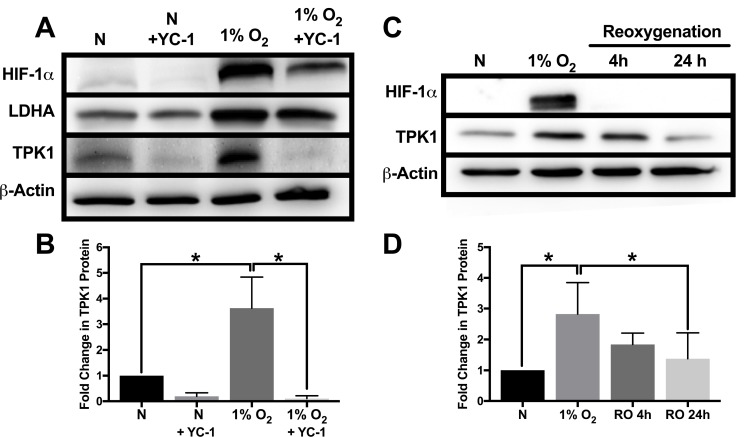
Figure 2. Attenuation of TPK1 expression using pharmacological inhibition of HIF-1α and reoxygenation.
(A) Representative Western blots demonstrating HIF-1α, LDHA, and TPK1 protein expression under normoxic (N) and hypoxic conditions ± YC-1 in WCLs isolated from wild type HCT 116 cells seeded at 2500 cells/cm2 and pre-treated with 5 μM YC-1 for 24 h prior to 48 h hypoxic exposure in the presence of 5 μM YC-1. β-Actin expression serves as the loading control. (B) Densitometry analysis of the fold change in TPK1 expression ± SD following exposure to 1% O2 in the presence or absence of YC-1 for wildtype HCT 116 cells compared to untreated normoxic control (N) including n = 3 independent experiments. (C) Representative Western blots demonstrating HIF-1α and TPK1 in WCLs isolated from wild type HCT 116 cells seeded at 1250 cells/cm2 and treated in 1% O2 for 48 h with subsequent reoxygenation at 21% O2 for 4 and 24 h. (D) Densitometry analysis of the fold change in TPK1 expression ± SD following exposure to 1% O2 and subsequent reoxygenation in wildtype HCT 116 cells compared to untreated normoxic control (N) including n = 4 independent experiments. (*) Represents statistically significant difference (p < 0.05) based on results of one-way ANOVA with Tukey’s post-hoc test.

