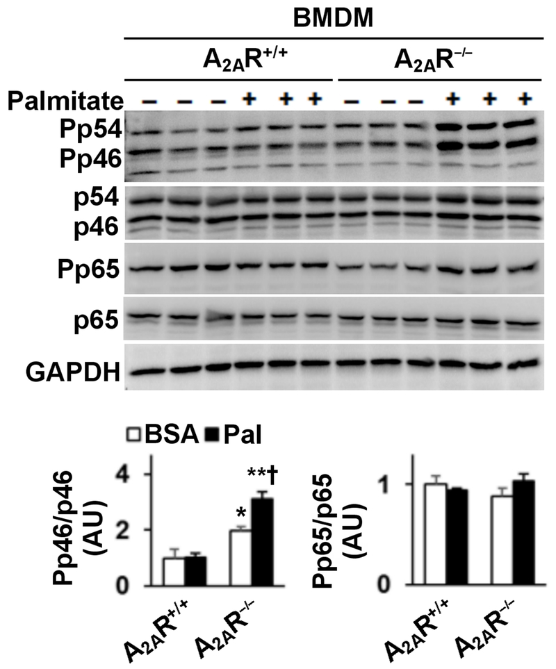
Figure 6. A2AR disruption enhances macrophage proinflammatory signaling.
Bone marrow cells were isolated from male A2AR−/− and A2AR+/+ mice, and differentiated into macrophages (BMDM). Cells were incubated in growth media in the presence of palmitate (250 µM, conjugated in BSA) or BSA for 24 hr. Cell lysates were subjected to Western blot analysis. Bar graphs, quantifications of blots. Numeric data are means ± SEM. n = 6 - 8. Statistical difference between A2AR−/− and A2AR+/+ with the same treatment: **, P < 0.01.

