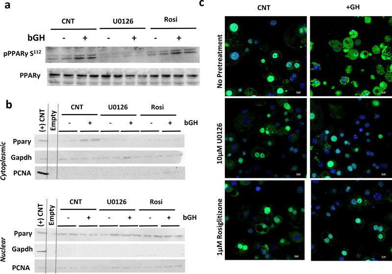
Figure 3. GH Treatment Results in Rapid PPARγ Translocation.
a) Representative Western blot analysis of pPPARγ S112 and total pPPARγ in 3T3-L1 adipocytes treated with 500ng/mL bGH for 20minutes after 2 hours of pre-treatment with 10 μM U0126 or 1 μM Rosiglitazone.
b) Representative Western blot analysis for PPARγ in 3T3-L1 adipocytes treated with 500ng/mL bGH for 1 hour after 2 hours of pre-treatment with MEK1 inhibitor, 10 μM U0126, or 1 μM Rosiglitazone following nuclear fractionation. The positive control is whole cell lysate of 3T3-L1 adipocytes. Gapdh and PCNA are loading controls for the cytoplasmic and nuclear fractions, respectively.
c) Immunofluorescence for PPARγ (green) with nuclei counterstained with DAPI (blue) of in 3T3-L1 adipocytes treated with 500ng/mL bGH for 1 hour after 2 hours pre-treatment with 10 μM U0126 or 1 μM Rosiglitazone.

