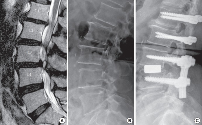
Fig. 4.

The images of a 66-year-old female patient. (A) Preoperative magnetic resonance imaging T2-weighted image sagittal view showed herniated and migration of intervertebral disc at L1–2–3–4. Spondylolisthesis was noted at L3–4. Preoperative (B) and postoperative (C) radiographies demonstrated well-maintained lumbar lordosis.
