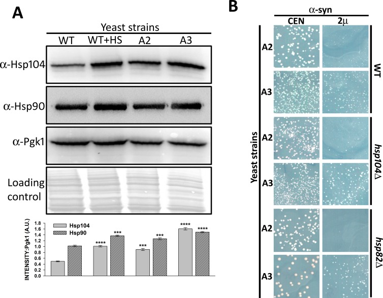
Fig 3. Reduction in α-syn toxicity in strain A3 was independent of elevated Hsp104 or Hsp90 levels.
WT, A2, or A3 cells were grown in liquid media until mid-log phase. For heat shock, wt cells were incubated at 37°C for 30 minutes. (A) The whole-cell lysates were probed with anti-Hsp104, anti-Hsp90, or anti-Pgk1 (loading control). As seen, strain A3 showed elevated levels of Hsp90 and Hsp104, which was similar to wt cells following heat shock treatment. The bottom panel shows quantification of Hsp104 and Hsp90 levels, normalized to Pgk1. (B) The indicated strains were transformed with either CEN or 2µ plasmids encoding α-syn under the strong constitutive GPD promoter. Growth phenotypes were monitored after 3 days at 30°C and 2 days at 25°C. Averages from a minimum of three independent experiments are presented. Error bars represent the standard error. P-values were calculated using paired t-test using wt as a control.

