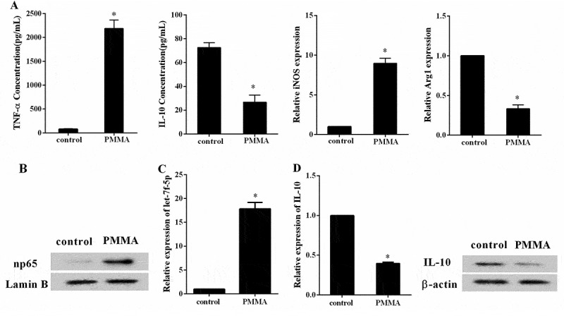
Figure 2.

The expression of let-7f-5p was increased in the process of M1 macrophage polarization that induced by PMMA. RAW264.7 cells were assigned into control and PMMA group. (a) The concentration of TNF-α and IL-10 in RAW264.7 cells was detected by ELISA, and the expression of iNOS and Arg1 in RAW264.7 cells was quantified by qRT-PCR. *P < 0.05 vs. control. (b) The expression of np65 and Lamin B protein in RAW264.7 cells was analyzed by western blot. (c) The expression of let-7f-5p in RAW264.7 cells was determined by qRT-PCR. *P < 0.05 vs. control. (d) The expression of IL-10 mRNA and protein in RAW264.7 cells was determined by qRT-PCR and western blot, respectively. *P < 0.05 vs. control.
