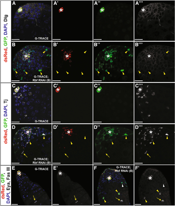
Figure 2. Hub Cells Convert to Cyst Lineage Cells upon Rbf Knockdown.
(A–F) Single confocal sections through the testis apex of E132ts > G-TRACE control testes (A, C, and E) and E132ts > G-TRACE; Rbf RNAi experimental testes (B, D, and F). Marking and RNAi induction occurred for 7 days (A–D) or 14 days (E and F) at 29°C. Testes are immunostained for dsRed (Gal4-expressing hub cells; red), GFP (hub cells and cells derived from hub cells; green), DAPI (nuclei; blue), and either Dlg (cell membranes; white, A and B), Tj (cyst lineage; white, C and D), or Fas III and Eya (hub membrane and late cyst cells, respectively; white, E and F). Merged and single channels are shown.
(A–Aʹʹʹ) Hub cells (white asterisk) are seen in a confined area at the apical tip of the control testis. These cells express the hub-GAL4 driver, allowing continuous expression of both RFP and GFP, causing cells to appear yellow. Merged (A), dsRed only (Aʹ), GFP only (Aʹʹ), and Dlg only (Aʹʹʹ) channels are shown.
(B–Bʹʹʹ) Rbfknockdown causes hub cellconversion. Cells within the main hub cluster (white asterisk) still express the hub-GAL4 driver and appear yellow. Cells originating from the hub that have migrated away from the main hub cluster have lost their hub cell identity (yellow arrowheads) and appear only green. Merged (B), dsRed only (Bʹ), GFP only (Bʹʹ), and Dlg only (Bʹʹʹ) channels are shown.
(CCʹʹʹ) Cyst lineage cells (Tj; white) do not express either RFP or GFP and thus are not derived from converting hub cells in control testes. Merged (C), dsRed only (Cʹ), GFP only (Cʹʹ), and Tj only (Cʹʹʹ) channels are shown.
(D–Dʹʹʹ) Upon Rbf knockdown, hub-derived traced cells (RFP−GFP+) express the cyst lineage marker Tj (yellow arrowheads), indicating that these cells have changed their cell fate. Tj-positive cells not derived from the hub (RFP−GFP−) are also seen (yellow arrow). See also Figure S3. Merged (D), dsRed only (Dʹ), GFP only (Dʹʹ), and Tj only (Dʹʹʹ) channels are shown.
(E and Eʹ) Late cyst cells are unmarked (RFP−GFP−), express Eya (nuclear white; yellow arrow), and are seen far away from the hub (white asterisk) in control testes. Merged (E) and Eya and FasII only (Eʹ) channels are shown.
(F and Fʹ) Upon Rbf knockdown, some converted hub cells (RFP−GFP+) that are far away from the hub (white asterisk) are seen expressing the late cyst marker Eya (nuclear white; yellow arrowheads), indicating that they can progress through cyst lineage differentiation. Unmarked (RFP−GFP−) late cyst cells (Eya+) are also seen (yellow arrow). Some marked cells (RFP−GFP+) do not express Eya (white arrowhead), suggesting that these cells have lost hub fate but have not yet acquired later cyst cell fate. Merged (F) and Eya and FasII only (Fʹ) channels are shown.
Scale bars represent 20 μm. See also Figures S2–S4, Table S1, and Videos S1, S2, and S3.

