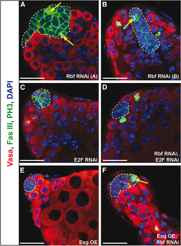
Figure 4. E2F Knockdown but Not Esg Overexpression Suppresses Hub Cell Proliferation Caused by Rbf Knockdown.
(A–F) Single confocal sections through the testis apex immunostained for Vasa (germ cells; red), Fas III (hub; membranous green), PH3 (mitotic cells; nuclear green), and DAPI (nuclei; blue). Hubs are outlined in white. Flies were shifted to 29°C for 14 days to induce RNAi knockdown using the E132ts driver.
(A and B) Testes with Rbf knocked down in the hub using two independent RNAi lines denoted A (A) and B (B) have extensive hub cell proliferation, indicated by PH3-marked hub cells (yellow arrows), leading to an enlargement of the hub.
(C and E) Knockdown of E2F (C) or overexpression of Esg (E) individually in the hub does not induce hub cell divisions, as indicated by the absence of PH3 in hub cells. Note that the hub size also appears normal.
(D) Knockdown of both E2F and Rbf in the hub suppresses hub cell proliferation as indicated by an absence of PH3 in the hub. Hub size appears normal. (F) Overexpression of Esg in the hub does not suppress the proliferation phenotype caused by Rbf knockdown. An enlarged hub with PH3-marked hub cells (yellow arrow) is still detected.
Scale bars represent 20 μm. See also Table 1.

