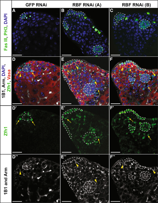
Figure 5. Loss of Rbf in Hub Cells Causes Ectopic Niche Formation.
(A–F) Single confocal sections through the testis apex of E132ts > GFP RNAi control testes (A and D) or E132ts > Rbf RNAi testes (B, C, E, and F). RNAi was induced for 14 days at 29°C. (A–C) Testes immunostained with Fas III (hub; membranous green), PH3 (mitotic cells; nuclear green), and DAPI (nuclei; blue). Control testis (A) contains only a single hub (white outline), while those testes expressing either Rbf RNAi A (B) or Rbf RNAi B (C) in hub cells contain multiple hubs. (D–F) Testes immunostained with Armadillo (hub; membranous white), 1B1 (fusome; white), Zfh1 (CySCs; green), Vasa (germ cells; red), and DAPI (nuclei; blue).
(D–Dʹʹ) Control testis with a single hub surrounded by both CySCs (yellow arrows) and GSCs represented by red cells with a dot fusome (yellow arrowhead). Merged (D), Zfh1 only (Dʹ), and 1B1 and Arm only (Dʹʹ) channels are shown.
(E–Fʹʹ) Testes with Rbf knocked down in the hub using two independent RNAi lines denoted A (E–Eʹʹ) and B (F–Fʹʹ) contain multiple hubs (white outlines) scattered throughout the testis apex, each with CySCs (yellow arrows) and GSCs (yellow arrowheads). Merged (E and F), Zfh1 only (Eʹ and Fʹ), and 1B1 and Arm only (Eʹʹand Fʹʹ) channels are shown.
Scale bars represent 20 μm. See also Figure S5 and Video S4.

