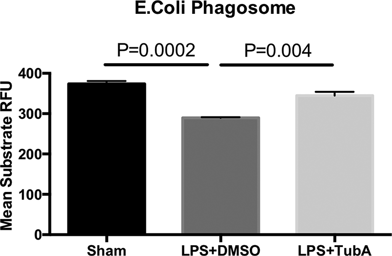
Fig. 4. Tubastatin A enhanced phagocytosis of RAW264.7 in vitro.

Macrophages were treated at 37 °C in the absence or presence of LPS (1μg/mL) or Tubastatin A (10 μM) for 3 h, then incubated with 1 mg/ml pHrodo E. coli bioparticles and the fluorescence analyzed on a plate reader. Mean values are relative fluorescence units (RFU), calculated from 3–5 wells per group subtracting the base-line fluorescence from wells containing pHrodo E. coli bioparticles but no cells (Means ± SEM, n = 3–5/group). LPS lipopolysaccharides, DMSO Dimethyl sulfoxide, TubA Tubastatin A, Sham no LPS, no treatment
