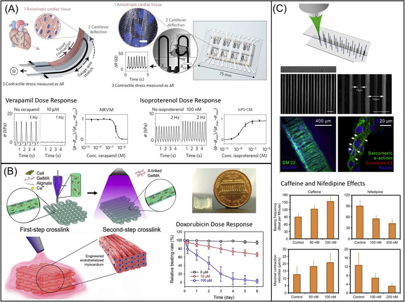
Fig. 3.
3D bioprinting of cardiac tissue models: (A) Schematic diagram showing the design of a multimaterial, patterned, piezoresistive stress sensor that aligns cardiomyocytes in a thick tissue and sense cardiac force output by changes in resistivity during contraction. Scale bar are 10 μm. Plots showing verapamil and isoproterenol dose response are on the bottom. (Reprinted from: [215]) (B) Schematic diagram showing the extrusion-based 3D printing system that generate multimaterial prints of cardiomyocytes and endothelial cells in naturally-based alginate and GelMA scaffold. The image of printed construct is shown on the top right and the doxorubicin dose response is shown on the lower right. (Reprinted from: [216]) (C) Schematic diagram showing TPP-based printing of micron-scale filaments, which was seeded with healthy and Long-QT iPSC-CMs. Fluorescence images in the middle showing the construct in bulk and cell alignment in various conditions. Bar charts on the bottom showing effects of caffeine and nifedipine on beating frequency and maximal contraction (Reprinted from: [217]).

