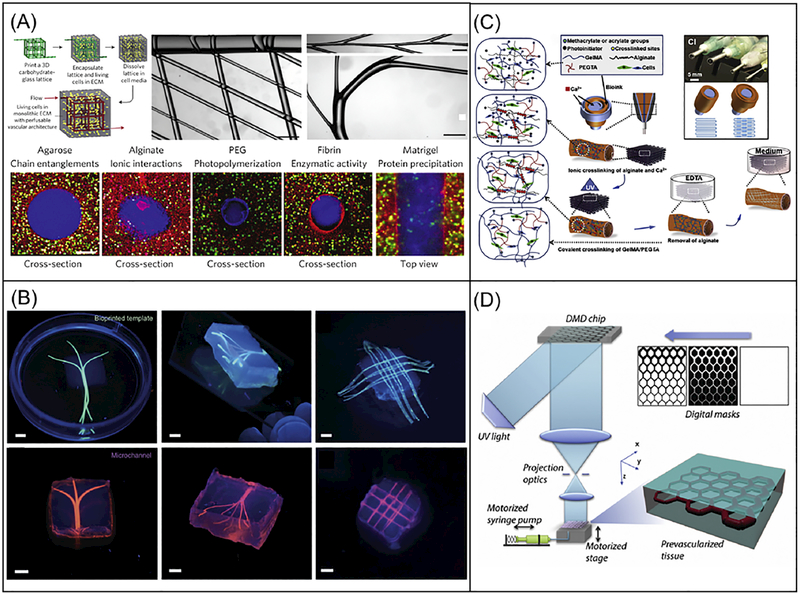
Fig. 4.
3D bioprinting of vascularized tissue models. (A) Schematic of a sacrificial bioprinting method using an extrusion-based bioprinter and carbohydrate glass as the sacrificial template, with images showing multiscale structures on top middle and right. Scale bars are 1mm. Bottom cross-sectional fluorescence images showing lumen structures from a variety of cell-laden ECM materials. Scale bars are 200 μm. (Reprinted from: [219]). (B) Fluorescence images showing the bioprinted alginate templates (green) enclosed in GelMA hydrogels and the respective microchannels perfused with a fluorescent microbead suspension (pink) after removal of the alginate templates. Scale bars are 3mm. (Reprinted from: [221]). (C) Schematic of the direct vasculature printing with a multilayered coaxial extrusion-based bioprinter and a blend bioink with two independent crosslinking mechanisms. (Reprinted from: [17]). (D) Schematic of the DLP-based bioprinting system for the rapid printing of prevascularized 3D tissues with direct encapsulation of endothelial and supportive cells in a continuous fashion. (Reprinted from: [127]).

