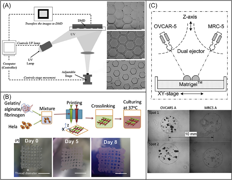
Fig. 5.
(A). Schematic diagram of a DLP-based system that uses a programmable DMD to selectively illuminate UV light onto photosensitive monomer solution. Bright-field images on the right showing HeLa cells-seeded PEGDA scaffolds with channels of 25-μm width (top), 45-μm width (middle), and 120-μm width (bottom). Scale bars are 100 μm. (Reprinted from: [240]). (B). Schematic process of an extrusion-based printing of gelatin/alginate/fibrinogen constructs with Hela cells to model cervical tumor. Bright field images on the bottom showing 3D printed Hela cell constructs on day 0, day 5 and day 8. Scale bar are 5 mm. (Reprinted from: [241]). (C). Schematic of an ejection printing platform composed of an automated stage and two nanoliter ejectors to dispense cancer cells (OVCAR-5) and fibroblasts (MRC-5). Bright-field image on the bottom showing 3D printed constructs with OVCAR-5 and MRC-5 cells. (Reprinted from: [149]).

