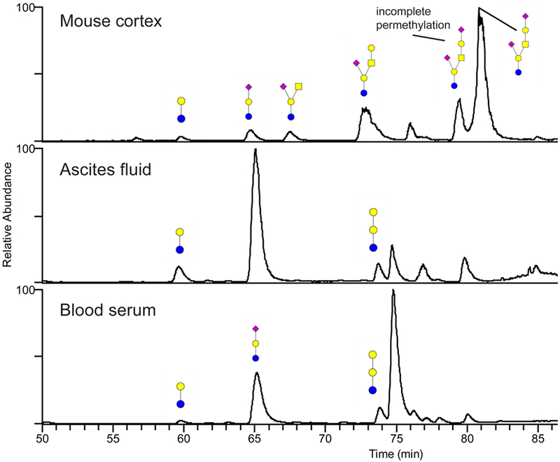
Figure 7.

Annotated base-peak chromatograms from the permethylated glycosphingolipid oligosaccharides released from mouse brain, human ascites fluid and serum. The glycans were separated on a porous graphitized carbon column maintained at 75°C prior to MS detection as protonated ions in the positive-ion mode. The annotation uses color cartoon nomenclature according to Varki et al. [57]. Some incompletely permethylated glycans were observed preceding the major peaks in the chromatogram, most markedly for the two peaks around 80 minutes in mouse cortex. The incompletely permethylated glycans were verified with MS/MS.
