Figure 1.
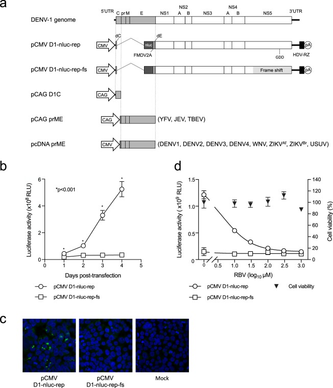
Construction and characterization of a DNA-based DENV1 replicon containing the NanoLuc gene. (a) Schematic representation of the DENV1 genome and replicon construct showing the position of the CMV promoter (CMV), NanoLuc gene (nluc), 2 A protein sequence of foot-and-mouth disease virus (FMDV2A), hepatitis delta virus ribozyme (HDV-RZ), and polyadenylation signal (pA). Structural protein-expression plasmids used to generate SRIPs are also shown. (b) Expression of reporter genes in DENV1 replicon. 293 T cells were transfected with pCMV-D1-nluc-rep or pCMV-D1-nluc-rep-fs, and luciferase activity was monitored at indicated time points. The mean and standard deviation calculated from triplicate NanoLuc values for each replicon are presented in the graph. The statistical significance of differences was evaluated using Student’s t-test. (c) dsRNA staining of cells transfected with replicon plasmid. 293T cells were transfected with the indicated plasmids and then stained with anti-dsRNA antibody (Green). Cell nuclei were counterstained with DAPI. (d) Luciferase activity of 293T cells transfected with pCMV-D1-nluc-rep in the presence of ribavirin at the indicated concentration. Detection was performed at 3 days post-transfection. Data are expressed as means of triplicate values with error bars indicating standard deviations.

