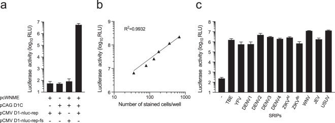
Figure 2.
Generation of multiple flavivirus SRIPs with different prME plasmids. (a) Luciferase activity of SRIPs produced by transfection of 293T cells with replicon plasmid and structural protein-expression plasmids. Each plasmid used for transfection is indicated. The supernatants of transfected cells collected at 3 days post-transfection were used to inoculate Vero cell monolayers. Luciferase activity of infected cells was subsequently determined at 3 days post-infection. (b) Relationship between luciferase activity and a number of infected cells. WNV-SRIPs were serially diluted (2-fold dilutions) and used to inoculate Vero cells. Luciferase activity and a number of cells stained with anti-NS1 antibody were plotted. The dotted line indicates the linear regression line. The coefficient of determination (R2) is displayed in the graph. (c) Luciferase activity of SRIPs produced by transfection of 293T cells with replicon plasmid and structural protein-expression plasmids. Each prME plasmid used for transfection is indicated. The supernatants of transfected cells collected at 3 days post-transfection were used to inoculate Vero cell monolayers. Luciferase activity of cells was subsequently determined at 3 days post-infection.

