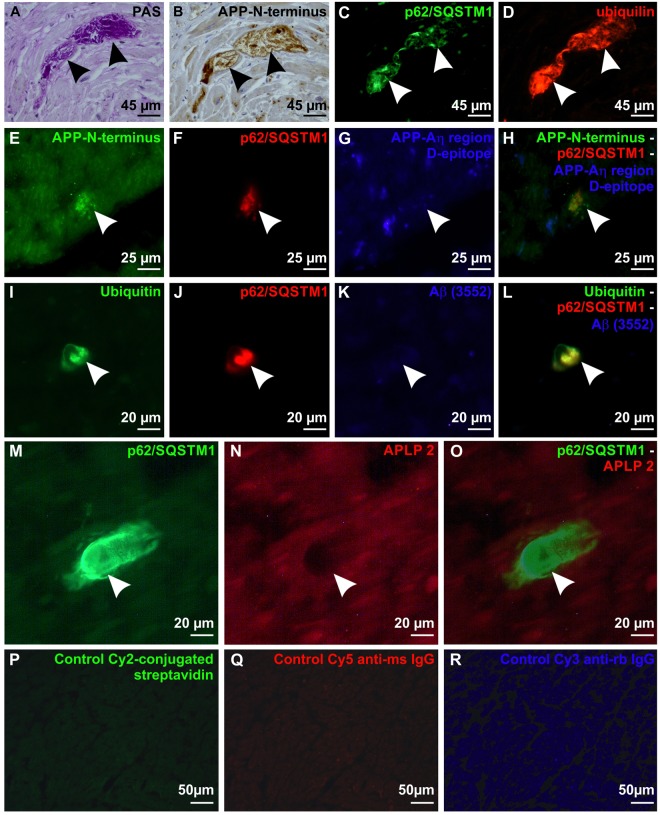
Figure 2.
Ubiquitinated and glycosylated p62/SQSTM1-immunopositive BD-inclusions exhibiting N-terminal epitopes of APP in cardiomyocytes in cross sections through the left ventricle wall. (A) BD-inclusion (arrows) stained with the PAS histochemical staining method in a cardiomyocyte of the left ventricular myocardium in case No. 53. (B) Subsequent section of the BD-inclusion of case No. 53 depicted in A immunostained with anti-APP (22C11) antibodies against the N-terminus of APP (arrows). Note that neighboring cardiomyocytes exhibit a physiological APP expression in the cytoplasm, focally associated with lipofuscin granules. (C) p62/SQSTM1- immunohistochemistry showed in another subsequent section of the BD-inclusion of case No. 53 depicted in (A,B) the presence of p62/SQSTM1 (arrows). The aggregates exhibited an amorphous structure and were sharply delineated. (D) The section shown in C was also stained for ubiquilin by double label immunofluorescence, which was colocalizing with p62/SQSTM1 in the BD-lesion (arrows). (E–H) Triple label immunofluorescence of a BD-lesion in the myocardium of the left ventricle of case No. 17. The BD-lesion was stained with anti-APP (22C11) detecting the N-terminus of APP (arrow in E,H) and p62/SQSTM1 (arrow in F,H) whereas the antibody against the D-epitope of the Aη-region did not detect this lesion (arrow in G,H). (I–L) Triple label immunofluorescence of a BD-lesion in case No. 50 shows expression of ubiquitin (arrow in I,L) and p62/SQSTM1 (arrow in J,L) but no expression of Aβ detected with the antibody (3552)44 (arrow in K,L). (M–O) Double label immunofluorescence of a BD-lesion in the left ventricular myocardium of case No. 56 showed a p62/SQSTM1-positive BD inclusion (arrowhead in M,O) which was not visible with the antibody against APLP2 (arrowhead in N,O) whereas the cardiomyocytes showed a mild physiological cytoplasmic staining of APLP2. (P–R) Negative controls (myocardium of the left ventricle from case No. 17) for immunofluorescence stainings were performed by omitting primary antibodies (only secondary antibodies/detection agents): Carbocyanin-2 (Cy2)-conjugated streptavidin (P) Cy5-conjugated anti-mouse IgG (Q) and Cy3-conjugated anti-rabbit IgG secondary antibodies (R). Positive controls are depicted in Suppl. Fig. 1.

