Figure 3.
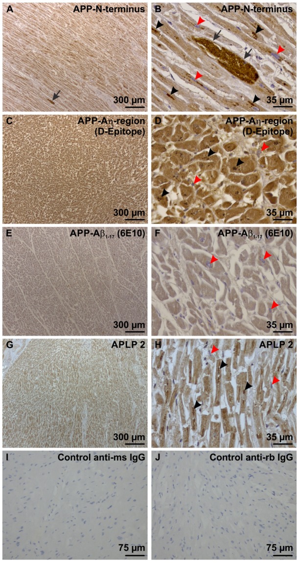
Physiological expression of APP and APLP2 in heart muscle cells of the left ventricle. (A) With an antibody directed against the N-terminus of APP there was a mild cytoplasmic staining in all cardiomyocytes of a left ventricle sample from case No. 15 exhibiting longitudinal-skewed cut muscle fibers. One BD lesion was seen (arrow) (B) In these cells lipofuscin particles (black arrowheads) were also stained as seen at the higher magnification level. The BD-inclusion (arrow) was morphologically different from the lipofuscin particles because it was bigger and showed a homogenous mass of strongly APP-positive material in the mildly APP expressing muscle cells. The nuclei of the cardiomyocytes were not stained and serve as intrinsic negative control for this staining (red arrowheads). (C) The Aη-D-epitope of APP was also expressed in all cardiomyocytes, here shown in a sample of the left ventricle myocardium of case No. 15. The muscle fibers were cut transversally. (D) At higher magnification level lipofuscin particles were also stained with the antibody against the Aη-D-Epitope (black arrowheads). BD-lesions were not seen with this antibody. The nuclei of the cardiomyocytes were not stained and served as intrinsic negative control for this staining (red arrowheads). (E,F) The Aβ epitope of APP was only faintly stained in cardiomyocytes of the left ventricle myocardium of case No. 58 using an antibody raised against Aβ1–17. No BD-lesions could be seen. Lipofuscin was not stained as well. The nuclei of the cardiomyocytes were not stained and served as intrinsic negative control for this staining (red arrowheads). The muscle fibers were cut transversally. (G) APLP2 was expressed in the cytoplasm of all cardiomyocytes of the left ventricular myocardium of case No. 1 exhibiting muscle fibers in longitudinal-skewed orientation. (H) At the higher magnification level lipofuscin particles (black arrowheads) were stained with this antibody as well. BD-lesions could not be detected with this antibody. The nuclei of the cardiomyocytes were not stained and served as intrinsic negative control for this staining (red arrowheads). (I,J) Negative controls by omitting the primary antibodies for biotinylated anti-mouse IgG (I) and anti-rabbit IgG secondary antibodies (J) were performed on left ventricular myocardium of case No. 44. Positive controls are shown in Suppl. Fig. 1.

