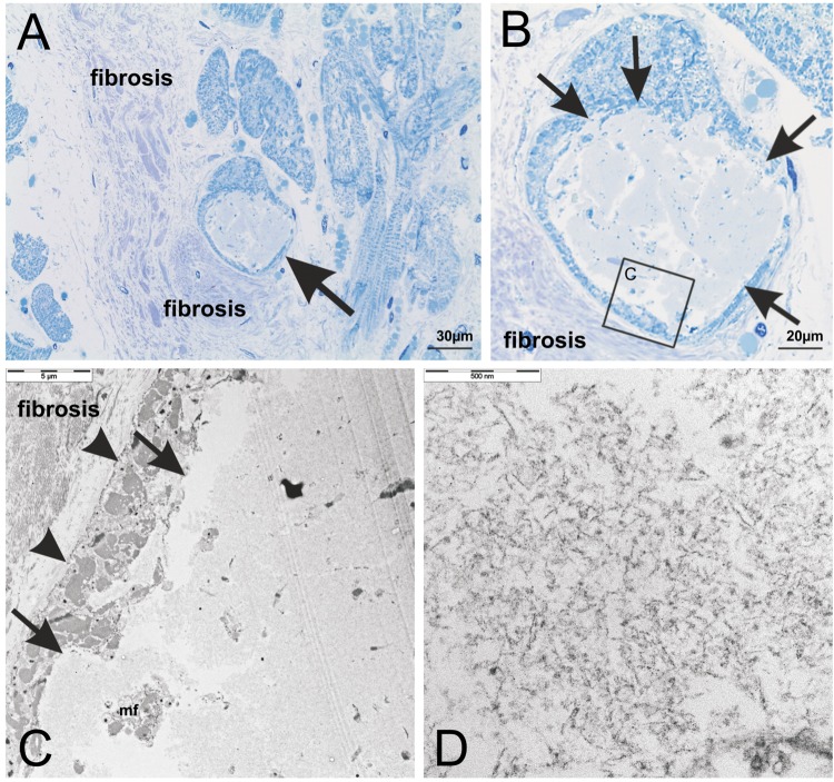
Figure 4.
(A) Structural analysis of cytoplasmic BD-inclusions in a semithin section of the left ventricular myocardium of case No. 53 stained with methylene blue. The inclusion body (arrow) was located intracellular in the sarcoplasm of a transversally cut cardiomyocyte and its morphological appearance identified this inclusion as BD-inclusion as described previously5. Cardiomyocytes were surrounded by cardiac fibrosis consisting of collagen fiber tissue. (B) Enlarged section of (A) The inclusion body was sharply delineated (arrows) and contained aggregates of amorphous material. The boxed area indicates the part of the cardiomyocyte depicted in C at the ultrastructural level. (C,D) The BD-inclusion was also analyzed in ultrathin sections by electron microscopy. The aggregates were not membrane-coated and sharply demarcated from surrounding myofibrils (C: arrows). Between the aggregates only few myofibrils (C: mf) and no preserved cell organelles (D) could be seen. The aggregated material exhibited a fibrillar pattern at the higher magnification level (D). The cell membrane of the cardiomyocyte was intact (C: arrow heads) and surrounded by cardiac fibrosis. This ultrastructural pattern confirmed these lesions to represent BD.

