Figure 5.
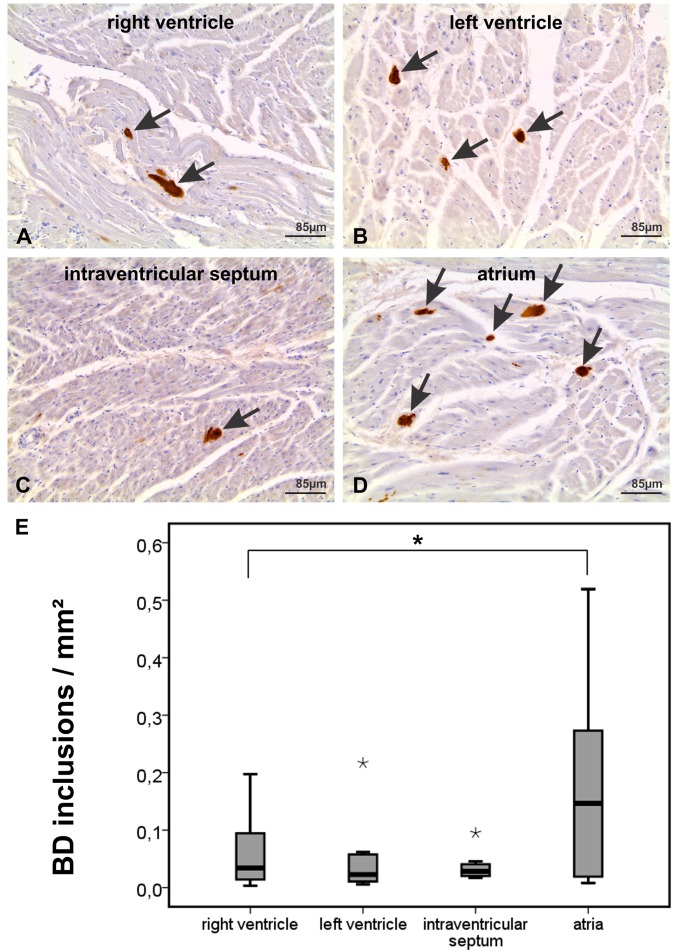
p62/SQSTM1-immunopositive BD inclusions were found in muscle cells of all parts of the heart of case No. 21 shown in transversal sections of the atrium, interventricular septum, and the left and right ventricles. (A) Two p62/SQSTM1-positive BD-inclusions are seen in this figure of the right ventricle (arrows). (B) Three BD-inclusions are marked with anti-p62/SQSTM1 in this image of the left ventricle (arrows). (C) The intraventricular septum image shows one BD-lesion (arrow). (D) The section of the atrium depicts five p62/SQSTM1-positive BD-lesions (arrows) indicating the high frequency of BD-lesions that could be detected in the atria by immunohistochemistry. The lesions were disseminated over the area of the atrial myocardium. (E) Assessment of the frequency of p62/SQSTM1-positive BD-inclusions in immunostained gross sections of the atria, ventricles and the intraventricular septum in cases No. 17, 21, 30, 33, 39, 44, 52, and 55 revealed the highest amount of inclusions in the atria and the lowest in the interventricular septum. The boxplot diagram shows the distribution of BD-inclusions/mm2 in the right and left ventricle, the interventricular septum and the atria (*statistical outliers, *significant at the 0.05 level (single sided Wilcoxon rank sum test), n = 8). The negative staining control showing only the staining with the secondary antibody while omitting the primary one is shown in Fig. 3 (I). The positive control for the p62/SQSTM1 antibody is depicted in Suppl. Fig. 1A.

