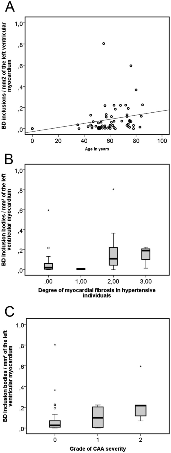
Figure 6.

The frequency of p62/SQSTM1-immunopositive BD-inclusions/mm2 of the left ventricular myocardium was compared to age (A) the degree of myocardial fibrosis in hypertensive individuals (B) and CAA severity (C). (A) The frequency of inclusions increased very mildly in correlation with the age as demonstrated in a scatter diagram (Linear regression analysis: R² = 0.074; β = 0.273, p = 0.032, n = 62, Table 2) (black line = regression line). (B) An association of p62/SQSTM1-positive BD-inclusions of the left ventricle with the degree of myocardial fibrosis in hypertensive individuals is shown in this boxplot diagram (Linear regression analysis controlled for age and gender: R² = 0.163; β = 0.302, p = 0.032, n = 62, Table 2). (C) The boxplot diagram shows the association between the frequency of left ventricular BD-inclusions and the CAA severity (Linear regression analysis controlled for age and gender: R² = 0.16; β = 0.273, p = 0.035, n = 62, Table 2). (B,C) ° and *statistical outliers).
