Figure 1.
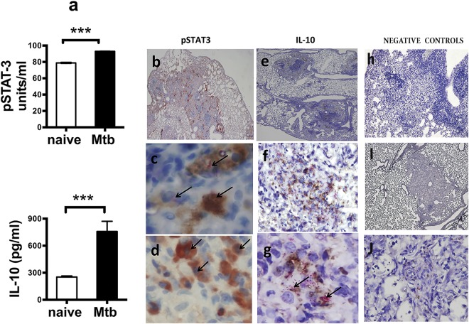
Elevated expression of pSTAT3 and IL-10 in the lungs of mice naïve and chronically infected with Mtb. (a) Lung homogenates obtained from naïve (white bars) or chronically Mtb infected (black bars) C57BL/6 mice (n = 5) were assayed by ELISA for pSTAT3 or by CBA for IL-10 to compare the levels of expression between groups of mice. Data represent mean ± SEM where ***p < 0.001. (b–j) Are photographs of lung tissue sections obtained from mice at 60 days of Mtb infection and stained by IHC only (b,d,h,i) or by IHC and acid fast staining (e,c,f,g,j). (b) pSTAT3 (red) positive staining in lesions, perivascular and bronchiolar cellular cuffs (original magnification 4×). (c) Co-localization of acid fast positive staining (fuchsia color with arrows) with positive staining for pSTAT3 (brown) in macrophage cells within the granuloma lesion (original magnification100×). (d) Positive staining for pSTAT3 (brown) in nuclei of macrophage cells within the granuloma lesion (original magnification 100×). (e) IL-10 (brown) within lesions of granulomas (original magnification 4×). (f,g) Co-localization of acid fast positive staining (fuchsia color) with positive staining for IL-10 (brown) in macrophages within the granuloma lesion (f) original magnification 40× and (g) original magnification 100×. (h) Negative control for pSTAT3 staining in which the first antibody was omitted (original magnification 10×) (i,j) negative control for IL-10 staining in which first antibody was omitted [I and J original magnification 10× and 100× respectively].

