Figure 4.
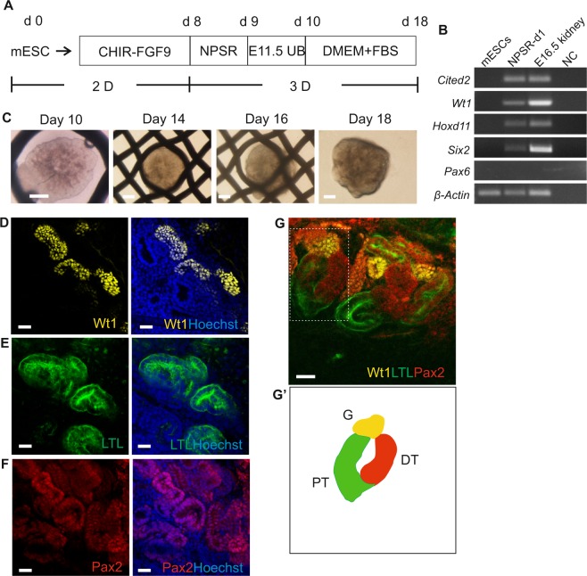
Generation of kidney organoids through aggregation of mESC-derived nephron progenitor cells and embryonic UB. (A) Schematic of the differentiation protocol of mESCs into kidney organoids. (B) Electrophoresis gel of RT-PCR products presenting kidney lineage cells expressing nephron progenitor markers Cited2, Wt1, Hoxd11, and Six2 after incubation in NPSR medium overnight. (C) Global bright field images of kidney organoids in Trowel culture. Scale bars: 500 μm. (D–F) Whole-mount immunofluorescence analyses of the organoids showing nephron progenitor markers: (D) glomeruli marker - Wt1, (E) proximal tubule marker - LTL, (F) nephron marker - Pax2. Scale bars: 20μm. (G) Confocal image showing three compartments of segmented nephron, including the distal tubule (DT, Pax2 + LTL−), proximal tubule (PT, LTL+) and the glomerulus (G, Wt1+). Dotted box shows single nephron that was used to generate a schematic diagram in (G’). Scale bars: 20 μm.

