Fig. 3.
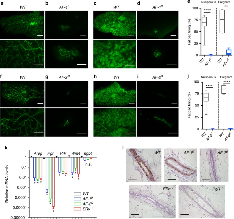
Mammary epithelial-intrinsic role of the ERα AF-1 and AF-2 domains. a–d Fluorescence stereomicrographs of contralateral inguinal mammary fat pads engrafted with mammary epithelium from AF-10.GFP+ or WT.GFP+ littermates. Nulliparous (a, b) and day 16–18 pregnant (c, d) recipients are shown. Scale bars; 5 mm (top), 2 mm (bottom). e Box plot showing extent of fat pad filling by the engrafted epithelia in virgin (n = 18) and pregnant (n = 5) recipients. f–j Fluorescence stereomicroscopy of contralateral inguinal mammary fat pads engrafted with mammary epithelium from AF-20.GFP+ or WT.GFP+ littermates. Nulliparous (f, g) and (P16–18) pregnant (h, i) recipients are shown. Scale bars; 5 mm (top) 2 mm (bottom). j Box plot showing extent of fat pad filling by the engrafted epithelia in virgin (n = 10) and pregnant (n = 4) recipients. For both box plots, horizontal lines outside the boxes depict minimum and maximum values, upper and lower borders of the box represent lower and upper quartiles and the line inside the box identifies the median. k Bar plot showing relative transcript levels of the ERα target genes Areg, Pgr1, Prlr and Wnt4, and a control gene, Itgb1, normalised to 36b4 and Hprt in mammary glands from peripubertal WT, AF-10, AF-20 and ERα−/− females. Data are shown as means ± SEM of three independent experiments. Paired two-tailed Student’s t test, *p < 0.05, **p < 0.01, ***p < 0.001, ****p < 0.0001, n.s. not significant. l PgR IHC of mammary glands from 3-week-old WT, AF-10, and AF-20, ERα−/− and PR−/− mice. Representative pictures of glands analysed from three females of each genotype are shown. Scale bar; 100 μm

