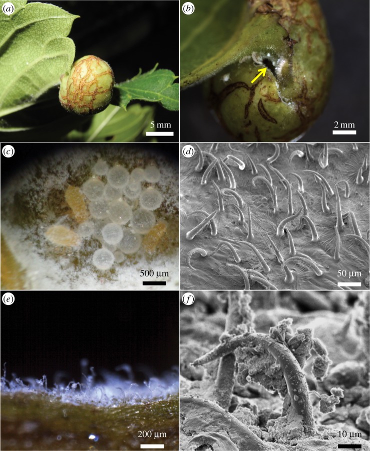
Figure 1.
Gall of C. clematis. (a) A mature gall on a leaf of Z. serrata. (b) A slit-like gall opening (arrow). (c) Young nymphs and honeydew balls in a mature gall. The gall inner cavity is full of aphid-derived powdery wax. (d) A scanning electron micrograph of trichomes on the gall inner surface. Note that aphid-derived wax is removed during fixation. (e) A fresh cross-section image of the gall inner surface. Note that trichomes are coated with aphid-derived white wax. (f) A scanning electron micrograph of the wax-coated trichomes. (Online version in colour.)

