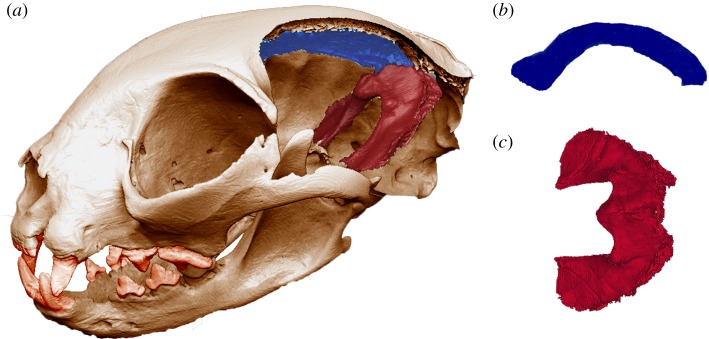
Figure 1.
(a) The skull used for the in silico model after performing a virtual parasagittal cut in the braincase to reveal the falx cerebri and the tentorium cerebelli (displayed in blue and red, respectively). (b) Falx cerebri in medial–lateral view. (c) Tentorium cerebelli in dorsal view. (Online version in colour.)

