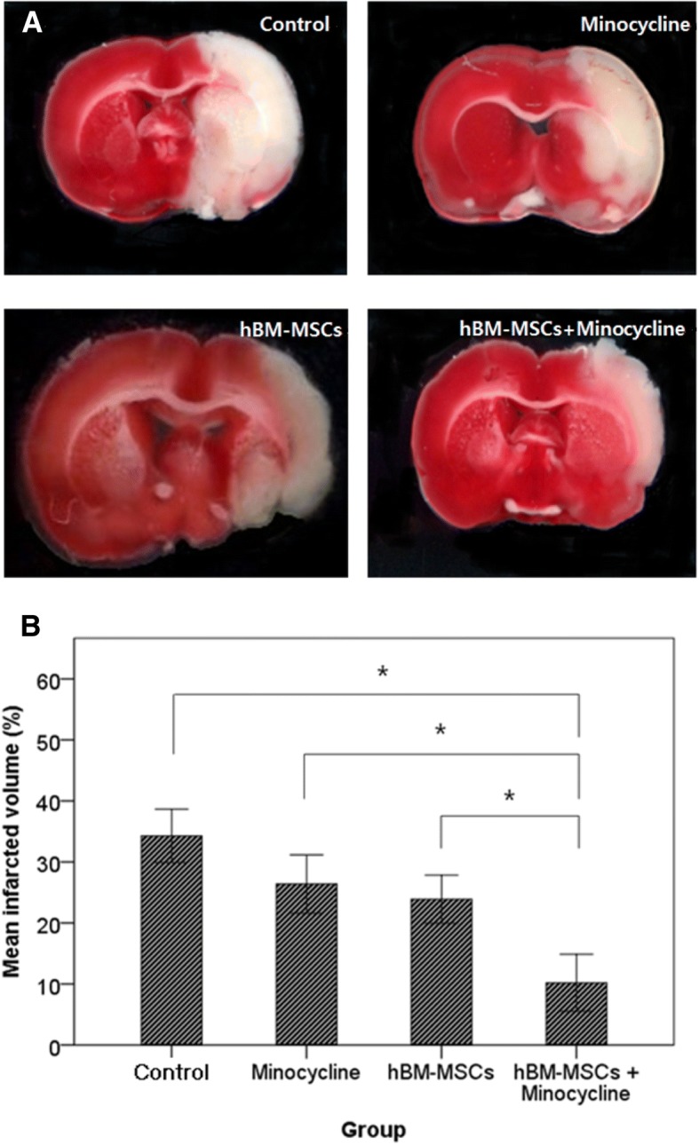
Fig. 4.

Volume of infarcted area. a Coronally sectioned brain tissue at a level of 10 mm from bregma with triphenyl tetrazolium chloride stain. b Quantitative analysis of infarction volume. The combination therapy group showed significantly lower infarction volume compared with other groups. Kruskal–Wallis test. *P <0.01. Abbreviation: hBM-MSC human bone marrow–derived mesenchymal stem cell
