Fig. 5.
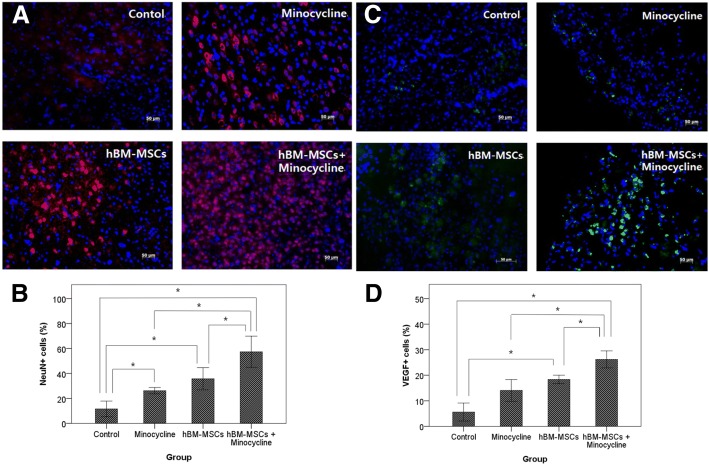
Neuronal nuclear antigen (NeuN) and vascular endothelial growth factor (VEGF) expression in the ischemic brain. a NeuN-positive cells in the ischemic boundary zone immunolabeled with red fluorescence. Scale bar = 50 um. c VEGF-positive cells in the ischemic boundary zone with immunolabeled green fluorescence. Nuclei were counterstained with 4–6-diamidino-2-phenyindole (DAPI) (blue). Scale bar = 50 um. b Quantitative analysis of NeuN-positive cells. d Quantitative analysis of VEGF-positive cells. Data are presented as mean. Kruskal–Wallis test, *P <0.01. Abbreviation: hBM-MSC human bone marrow–derived mesenchymal stem cell

