Figure 3.

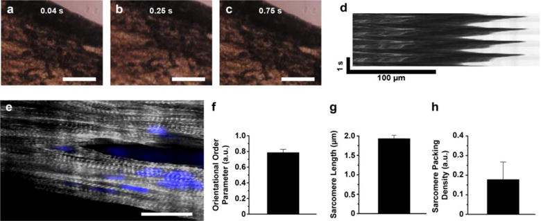
In vitro cardiomyocyte culture on nanofiber. a–d) (a–c) Bright field images of contraction of the fiber scaffolds with (d) kymograph. Scales of a-c are 100 μm. e) Confocal image of cardiomyocytes on nanofiber, stained for nuclei (blue) and α-actinin (grey). Scale is 20 μm. f–h) (f) Orientational order parameter (OOP), g) sarcomere length, and h) sarcomere packing density (SPD) of cardiomyocytes grown on the fibrous scaffolds. n=3 and ROI=4.
