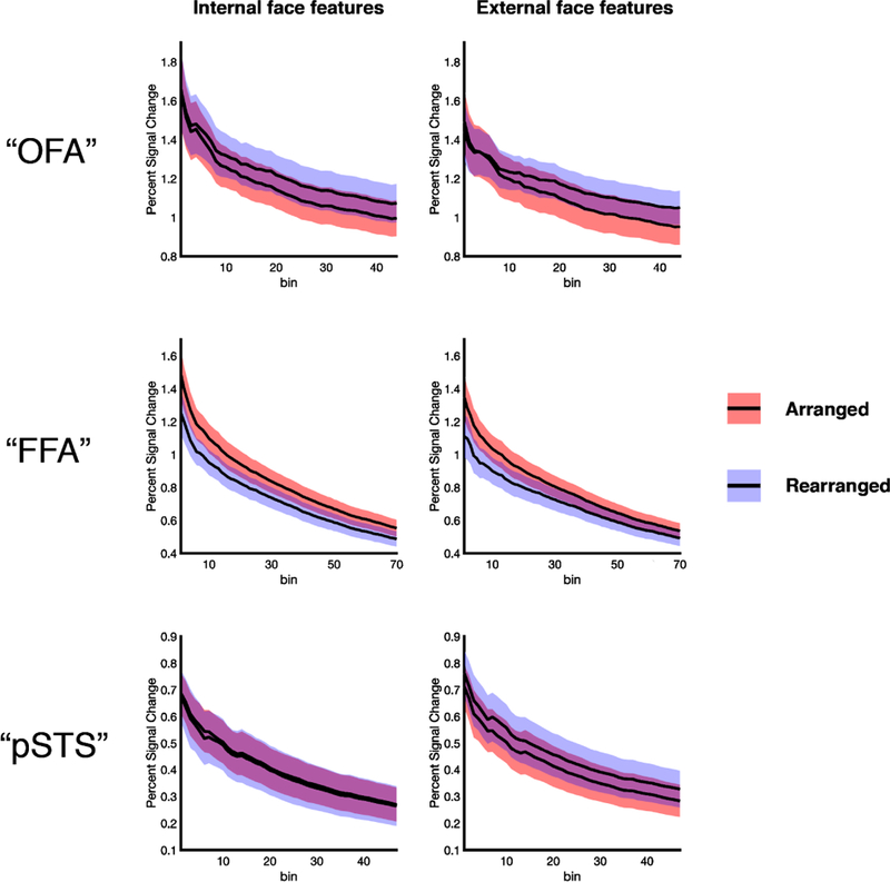
Figure 5.

VSF analysis exploring parts-based and whole-based representations in each face-selective region. The differential information processing performed across the three face-selective regions—with “OFA” and “pSTS” repres enting the parts of both internal and external of faces, and “FFA” representing the c oherent arrangement of both internal and external features—is found at every threshold u sed to define the ROIs. Colored bands around each line indicate the standard error of the mean.
