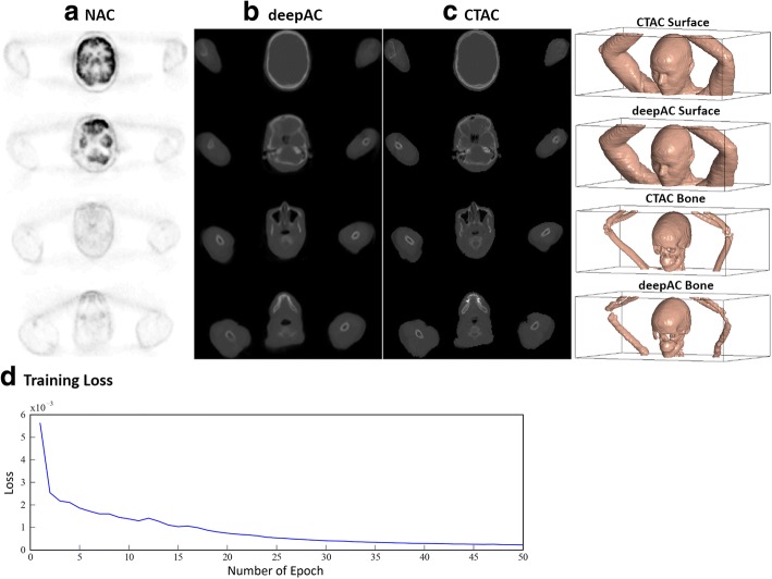
Fig. 3.
Example of pseudo-CT image from deepAC. Multiple axial slices from a the input NAC PET image, b the pseudo-CT generated using deepAC, and c the acquired CT. The 3D surface and bone model indicate a high similarity between the acquired CT and pseudo-CT. The surface and bone were rendered using a HU value of − 400 and 300, respectively. The training loss curve is shown in d

