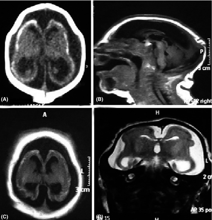
Figure 1.

Severe microcephaly, cerebral atrophy with ventriculomegaly, and prominent cerebrospinal fluid space. Axial CT scan (A) shows extensive, punctate cortico‐subcortical calcifications, cerebral atrophy, and bone overlap. Sagittal T1 MR image (B) shows craniofacial disproportion, occipital protuberance with redundant posterior skinfolds, and a hypoplastic corpus callosum. The fossa is relatively preserved. Axial T1 (C) and coronal T2 (D) demonstrate the abnormal gyral pattern with diffuse undersulcated cortex
