
Figure 2.
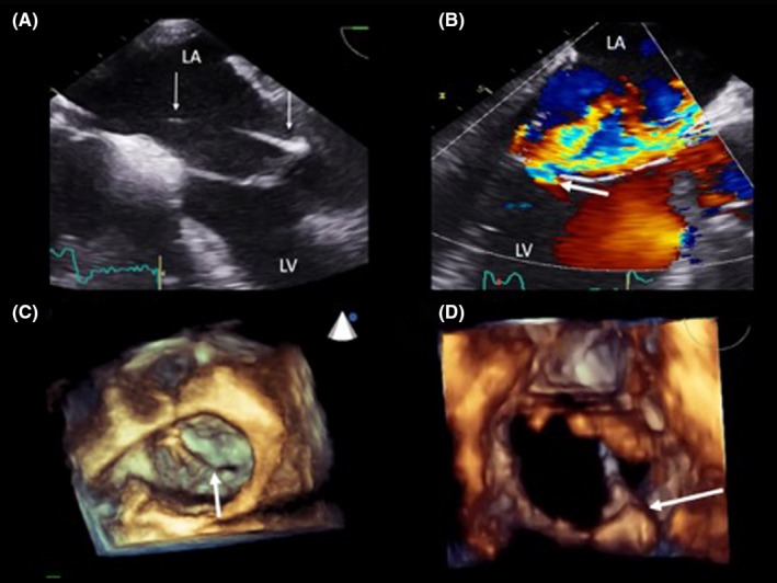
Trans‐esophageal echocardiogram (TEE). The LV lead is crossing the LA toward the posterior mitral valve leaflet at 0 degree (panel A). There is significant valvular regurgitation depicted by the white arrow at 120 degrees (panel B). Three‐dimensional reconstruction shows an en‐face view of the mitral valve with the white arrow demonstrating the endocardial LV lead preventing complete apposition of mitral valve leaflets at the P2/A2 scallops (panel C). The lead penetrates the mitral valve orifice at P2 (panel D)