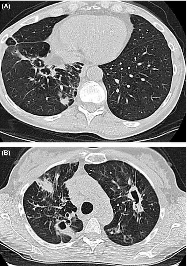
Figure 1.

A, Airspace consolidation within the right upper lobe measuring approximately 1.9 × 3.6 cm and increasing mediastinal lymphadenopathy. B, Progression with evidence of new cavitary lesion within the left upper lobe (lower scan) measuring 1.5 × 4.4 cm
