Fig. 2.
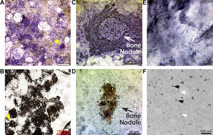
Membrane vesicles from mineralizing osteoblasts. A and B: mineralization and alkaline phosphatase in whole osteoblast cultures (). Mesenchymal stem cells were placed into culture as described in methods. After 3–4 wk, the cultures developed characteristic nodules of alkaline phosphatase and mineral-containing cells that can be seen directly in low power scans of cultures (1-mm fields). C and D: photomicrographs of representative bone nodules in osteoblast cultures. Low-power photomicrographs of nodules similar to the regions identified with arrows in A and B. Nodules of cells with strong alkaline phosphatase activity (C) and mineralization (D) are present. Only cultures with mineralizing osteoblasts had significant transport activity in isolated membranes (3, 21). E: oil immersion photomicrograph of osteoblast membranes labeled for alkaline phosphatase activity. Plasma membrane fragments were prepared by nitrogen cavitation in an isotonic HEPES-buffered sucrose solution (5). After alkaline phosphatase activity labeling, viewing at ×100 oil immersion revealed that the membrane fragments are alkaline phosphatase labeled, although the individual membranes are too small to resolve by light microscopy. F: electron microscopic analysis of osteoblast membrane fragments with alkaline phosphatase labeling. Examination by electron microscopy on methyl cellulose grids after alkaline phosphatase 20 nM gold labeling. About half of the membranes are labeled, consistent with alkaline phosphatase expression in apical but not basolateral membranes. Black arrows indicate labeled membrane fragments; white arrows indicate unlabeled membrane fragments.

